CASE20250608_001
When Old Stent Meets New Calcium
By Hing Yin Wilson Lam
Presenter
Hing Yin Wilson Lam
Authors
Hing Yin Wilson Lam1
Affiliation
Yan Chai Hospital, Hong Kong, China1
View Study Report
CASE20250608_001
Complex PCI - In-Stent Restenosis
When Old Stent Meets New Calcium
Hing Yin Wilson Lam1
Yan Chai Hospital, Hong Kong, China1
Clinical Information
Relevant Clinical History and Physical Exam
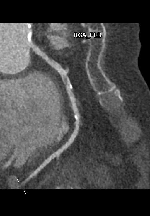
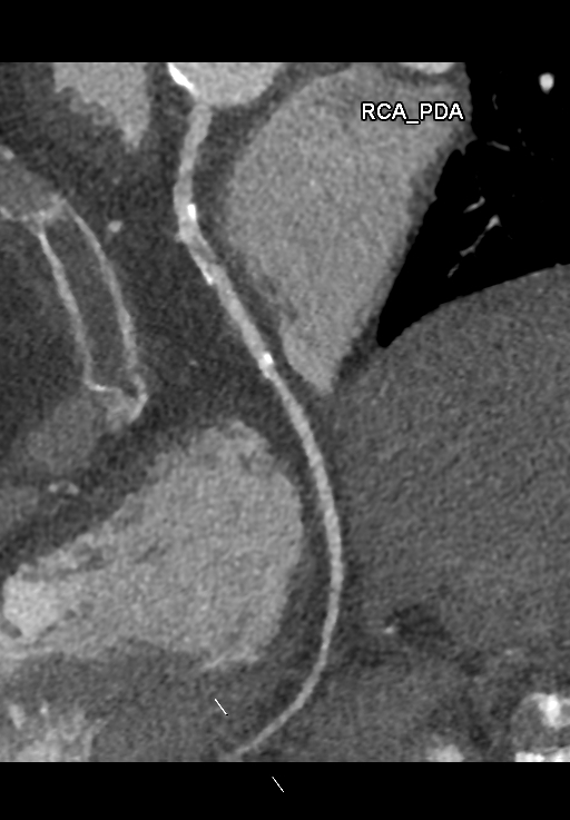
A 78-year-old woman with hypertension, hyperlipidemia, and a history of PCI to the right coronary artery with bare-metal stent placement in 2001 presented with a one-month history of stable angina. She underwent CT coronary angiography, which demonstrated moderate to severe in-stent restenosis in the RCA. She was admitted electively for coronary angiography and possible PCI. Physical examination was unremarkable, with no murmurs or signs of heart failure.
Relevant Test Results Prior to Catheterization
Laboratory investigations, including complete blood counts and liver and renal function tests, were within normal limits. Fasting glucose was 5.2 mmol/L, and LDL-C was 1.6 mmol/L.
Relevant Catheterization Findings
Dominance: Right
 COMPLEXPCI-1.mp4
COMPLEXPCI-2.mp4
Interventional Management
Procedural Step
The diagnostic angiogram confirmed significant ISR at the proximal RCA stent, with severe calcification, and a de novo significant stenosis in the mid-distal RCA.
COMPLEXPCI-1wire.mp4
COMPLEXPCI-2IVUS.mp4
COMPLEXPCI-3stent.mp4
Case Summary
This case illustrates the complex challenges associated with treating severe calcified in-stent restenosis in a long-standing bare-metal stent. The successful use of intravascular lithotripsy, supported by intravascular imaging, enabled effective lesion modification and optimal drug-eluting stent deployment. Contemporary interventional tools and imaging are essential for achieving safe and durable outcomes in complex coronary interventions.