
CASE20210724_002
Could We Simplify the Rescue for LM Complication?
By
Presenter
Antonia Anna Lukito
Authors
1
Affiliation
, Indonesia1
Complications - Complications
Could We Simplify the Rescue for LM Complication?
1
, Indonesia1
Clinical Information
Patient initials or Identifier Number
Mr YB
Relevant Clinical History and Physical Exam
A 59 yo male, presented with chest discomfort on exertion, came for second opinion, the exercise test was positive and coronary angiogram revealed two vessels disease were performed in another hospital.


Relevant Test Results Prior to Catheterization
The physical examination and laboratory were unremarkable.
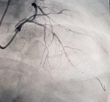
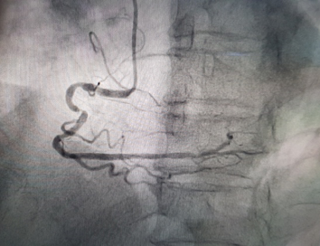
Relevant Catheterization Findings
The procedure via RRA, found LAD & RCA calcified lesions and high take-off ostial LCA. The dissection of mid LM run along the LAD, LCX and Intermediate was observed, after predilation of LAD. Patient developed back pain. A new GW inserted to LCX, while maintain the previous GW in LAD, and stenting the mid LAD and then stenting the ostial LM to proximal LAD, followed by postdilation and final POT at the ostial-mid LM.
Interventional Management
Procedural Step
The predilation was done using Across HP balloon 2.0-15mm at 6 to 10 atm along mid-distal LAD, then the dissection was observed at mid LM run along the LAD, LCX and Intermediate. Patient started complain of back pain. LM stenting was planned. The Sion blue GW inserted into LCX, while ensure not to lose the BMW guidewire in LAD, and started stenting the mid LAD using Xienxe Alpine DES 2.5-33mm at 8-10 atm and proceed the ostial LM to proximal LAD stenting using Yukon Chrome DES 3.5-16mm at 14 atm, followed by postdilation and final POT at the ostial-mid LM by NC Sprinter balloon 4.0-9mm at 8-10 atm. After stenting, the evaluation angio showed the resolved dissection at LM-LAD, the mild residual dissection was seen at Intermediate artery.
Case Summary
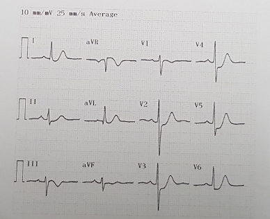
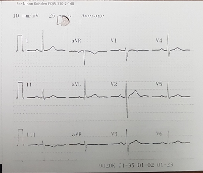
Patient went home two days later in stable condition, despite the new T wave-inversion on anterolateral leads.