
CASE20210819_004
Complete Percutaneous Coronary Revascularization of Calcified LM and TVD
By , , ,
Presenter
NAGENDRA BOOPATHY SENGUTTUVAN
Authors
1, 1, 1, 1
Affiliation
, India1
Complex PCI - Bifurcation/Left Main Diseases and Intervention
Complete Percutaneous Coronary Revascularization of Calcified LM and TVD
1, 1, 1, 1
, India1
Clinical Information
Patient initials or Identifier Number
2761994
Relevant Clinical History and Physical Exam
65 year old gentleman admitted with history of acute onset of chest pain which was retrosternal, compressive in nature, radiating to both shoulders. He was a known HTN. He was diagnosed with ACS (Anterior Wall Myocardial Infarction). Patient was lysed with 40mg inj. tenecteplase. Pain was reduced. He had one episode of ventricular tachycardia revert with antiarrythmic drug. Later posted for CAG.On physical examination vitals were within normal range. Systemic examination was normal.
Relevant Test Results Prior to Catheterization
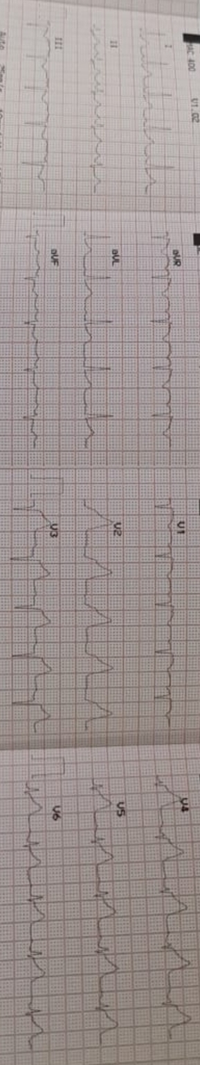
Hb- 16.5 g/dlPlatelets- 2.97 thousand/uLBUN- 10 mg/dlS.Creatinine- 0.7 mg/dlTroponin I - 9.91 ng/ml2D-ECHO- Mid anteroseptum, apical septum, LV apex, apical inferior wall, mid and apical anterior wall hypokinetic. Mild LV systolic dysfunction (EF- 45%) with grade I diastolic dysfunction. Mildly dilated LV, mild MR.ECG- ST elevation in V2-V6 with reciprocal changes in inferior leads.LDL- 226 mg/dl, TG- 239 mg/dl, S. Cholesterol- 327 mg/dlRecently diagnosed with DM II (HbA1c- 9.5%)
Relevant Catheterization Findings
Distal LM was 50% diseased.
 rca angio.mpg
se011 cag 2.mpg
se012 cag 3.mpg
Proximal LAD had 90% disease. Mid and distal segments had no flow limiting lesion. Branching vessels had non significant disease.
Proximal LCX had 90% lesion followed by long long lesion with maximum severity of 80% stenosis.
RCA was a dominant vessel with 90% focal lesion in mid segment.
Interventional Management
Procedural Step
-RCA hooked with 6F JR 3.5 guiding (Merit, USA)-Runthrough wire (Terumo, Japan) used to cross the lesion-Pre dilatation done with 1.5x10mm SC (Terumo, Japan) & 2.5x12mm NC balloons (Terumo, Japan)-Lesion stented with 3.5x20mm DES (Boston scientific, USA )-Post dilatation done with 3.0x8mm NC balloon(Terumo, Japan)). TIMI III flow achieved-Left coronary hooked with 7F BL 3.5 catheter (Terumo, Japan)-Runthrough wire (Terumo, Japan) and Fielder FC wire (Asahi, Japan) were crossed in LAD & LCX respectively-Predilatation in LCX done with 3.0x8mm NC balloon (Terumo, Japan) and in LAD with 3.0x10 NC balloon (Brosmed, China).-3.0x10mm cutting balloon (Brosmed, China) used to predilate proximal LCX and from LM to LCX-Using 3.0x20mm DES (Boston scientific, USA) ostioproximal LCX stented.-Using step crush technique with 3.0x10mm NC balloon (Brosmed, China) in LAD proximal struts of LCX stent were crushed.-LM to LAD stented using 3.0x34 mm DES (Medtronic, USA). Distal displacement of stent noted while deploying it.-LCX rewired and ostium was post dilated with 1.5x10mm SC balloon (Terumo, Japan)-Post dilatation in LAD & LCX done using 3.0x10mm NC (Brosmed, China) & 3.0X8mm NC (Terumo, Japan) respectively.-KBI done using same balloons used for post dilatation in LAD & LCX.-Ostial LM was stented using 4x8mm DES (Medtronic, USA) followed by post dilatation and aortic flurrying with stent balloon only. - TIMI III flow achieved- IVUS done pre and post stenting
rca pre dil.mpg
rca stented.mpg
se007 post dil.mpg
se009 rca final shot.mpg
se013 lcx predil with ...mpg
se014 lm to oLAD predil.mpg
se018 LCX pre dil 2-2.mpg
se021 lm to oLCX PREDIL.mpg
LCX POST BALLOON.wmv
se033 stent in lcx and balloon in lad.mpg
se034 lad balloon infl foll by stent infla in lcx.mpg
se041.mpg
se043 lcx ostium post dil with 1.5 balloon.mpg
se045 lcx post dil.mpg
se047 lad post dil.mpg
se048 KBI.mpg
se050.mpg
se052 oLM STENTING.mpg
se053 LM STENTING.mpg
se054 LM POST DIL.mpg
se055 AORTIC FLURING.mpg
se060final angio.mpg
Case Summary
- Complete revascularization can be done in stable ACS patients- Never miss left main ostium in LM stenting- Cutting balloon should be used for fibrotic lesions- Advise patient to avoid deep breathing during ostial stenting as it may displace the stent while deploying- Always try to use IVUS in LM and bifurcation lesions for better assessment of disease - Planned 2 stent technique should be preferred over provisional stenting in good size vessels