
CASE20210828_005
Troubleshooting with IVUS
By
Presenter
Jonathan Fang
Authors
1
Affiliation
, Hong Kong, China1
Imaging - Invasive Imaging (IVUS, OCT, spectroscopy, etc)
Troubleshooting with IVUS
1
, Hong Kong, China1
Clinical Information
Patient initials or Identifier Number
KCM
Relevant Clinical History and Physical Exam
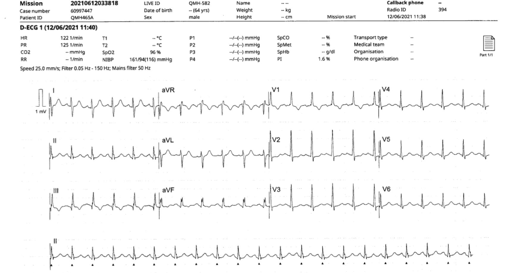
64 year-old man with diabetes, hypertension. Presents with chest pain for 2 days with new-onset CCS class III-IV symptoms No ankle edema, JVP not elevated on examination Killip class I Mildly tachycardiac with HR ~105bpm on examination
Relevant Test Results Prior to Catheterization
ECG V1-V4 TWI STD Echo EF 60% Troponin elevated Treated as NSTEMI
Relevant Catheterization Findings
Coro LM: NormalLAD: Heavily calcified (eccentric), proximal LAD moderate disease, mLAD 99% stenosisLCX: Large dominant vessel; LPDA 50% disease, large OM branch 40% diseaseRCA: Small non-dominant vessel
 MOVIE-0081.mp4
MOVIE-0082.mp4
MOVIE-0083.mp4
MOVIE-0084.mp4
MOVIE-0085.mp4
MOVIE-0086.mp4
Interventional Management
Procedural Step
Right radial access Guide: 6Fr EBU3.5PCI to LAD Wires: Sion Blue to LAD, Runthrough HC to D2Pre-dilatation: 2.5x12mm Euphora; IVUS performed for procedural planningFurther dilatation with 2.5x10mm NC Scoreflex at 20atm, 3.0x15mm NC Scoreflex at 20atm.Stent: 2.5x48mm Xience Sierra DES to mLADPatient complained of chest pain and ECG change noted. Loss of D2, rewired with Fielder XTA and dilatation with 1.5x10mm Ryurei balloon to D2 ostium; wiredescalated to Runthrough HCPost-dilatation: 2.5x12mm Accuforce at 20atm, 3.0x15mm NC Euphora at 20atm, 3.25x15mm Accuforceat 20atm, 3.75x12mm NC Trek at 12atmHowever patient still complained of chest pain on table IVUS showed intramural haematoma extending proximally to prox LAD. Review of angiogram showed rim of staining in proximal LAD with narrowing in pLAD corresponding to the location of haematoma Proximal LAD further stented with 3.25x23mm Xience Sierra DESPost-dilatation with 4.0x12mm NC Emerge. Chest pain subsided Good angiographic and IVUS results, TIMI 3 flow
MOVIE-0086.mp4
MOVIE-0087.mp4
MOVIE-0088.mp4
MOVIE-0089.mp4
MOVIE-0090.mp4
MOVIE-0091.mp4
MOVIE-0092.mp4
MOVIE-0093.mp4
MOVIE-0094.mp4
MOVIE-0095.mp4
MOVIE-0096.mp4
MOVIE-0097.mp4
MOVIE-0098.mp4
MOVIE-0099.mp4
MOVIE-0100.mp4
MOVIE-0101.mp4
MOVIE-0102.mp4
MOVIE-0103.mp4
MOVIE-0104.mp4
MOVIE-0105.mp4
MOVIE-0106.mp4
MOVIE-0107.mp4
MOVIE-0108.mp4
MOVIE-0109.mp4
MOVIE-0110.mp4
MOVIE-0111.mp4
MOVIE-0112.mp4
MOVIE-0113.mp4
MOVIE-0114.mp4
MOVIE-0115.mp4
MOVIE-0116.mp4
MOVIE-0117.mp4
MOVIE-0118.mp4
Case Summary
Intravascular imaging not only helps in procedural planning with determining sizing and the length of balloons and stents required. It also helps in troubleshooting potential complications. Imaging should be considered when there is uncertain on angiogram.