CASE20220806_001

"Hanging By A Thread" - A Challenging Case of Acute Coronary Syndrome caused by Critically Stenosed Distal Left Main and critically stenosis Right Coronary Artery due to Eruptive Calcified Nodules in an Octogenerian Patient.

By ,
like off

Presenter

Tjen Jhung Lee

Authors

1, 1

Affiliation

, Malaysia1
Complex PCI - Calcified Lesion

"Hanging By A Thread" - A Challenging Case of Acute Coronary Syndrome caused by Critically Stenosed Distal Left Main and critically stenosis Right Coronary Artery due to Eruptive Calcified Nodules in an Octogenerian Patient.

1, 1

, Malaysia1

Clinical Information

Patient initials or Identifier Number

461567

Relevant Clinical History and Physical Exam

We detailthe history of an 88 year old fit man withbackground hypertension and benignprostatic hyperplasia, planned for surgical treatment. A pre-operative Exercise stress ECHOshowed ST depressions with hypokinesia at the anterior segments at peakexercise.Invasivecoronary angiogram at another academic-based hospital showed severe calcified3VD thus he was advised for CABG but patient not keen due to the age-relatedrisks. He had chest pain and was readmitted for NSTEMI 2 weeks later.

Relevant Test Results Prior to Catheterization

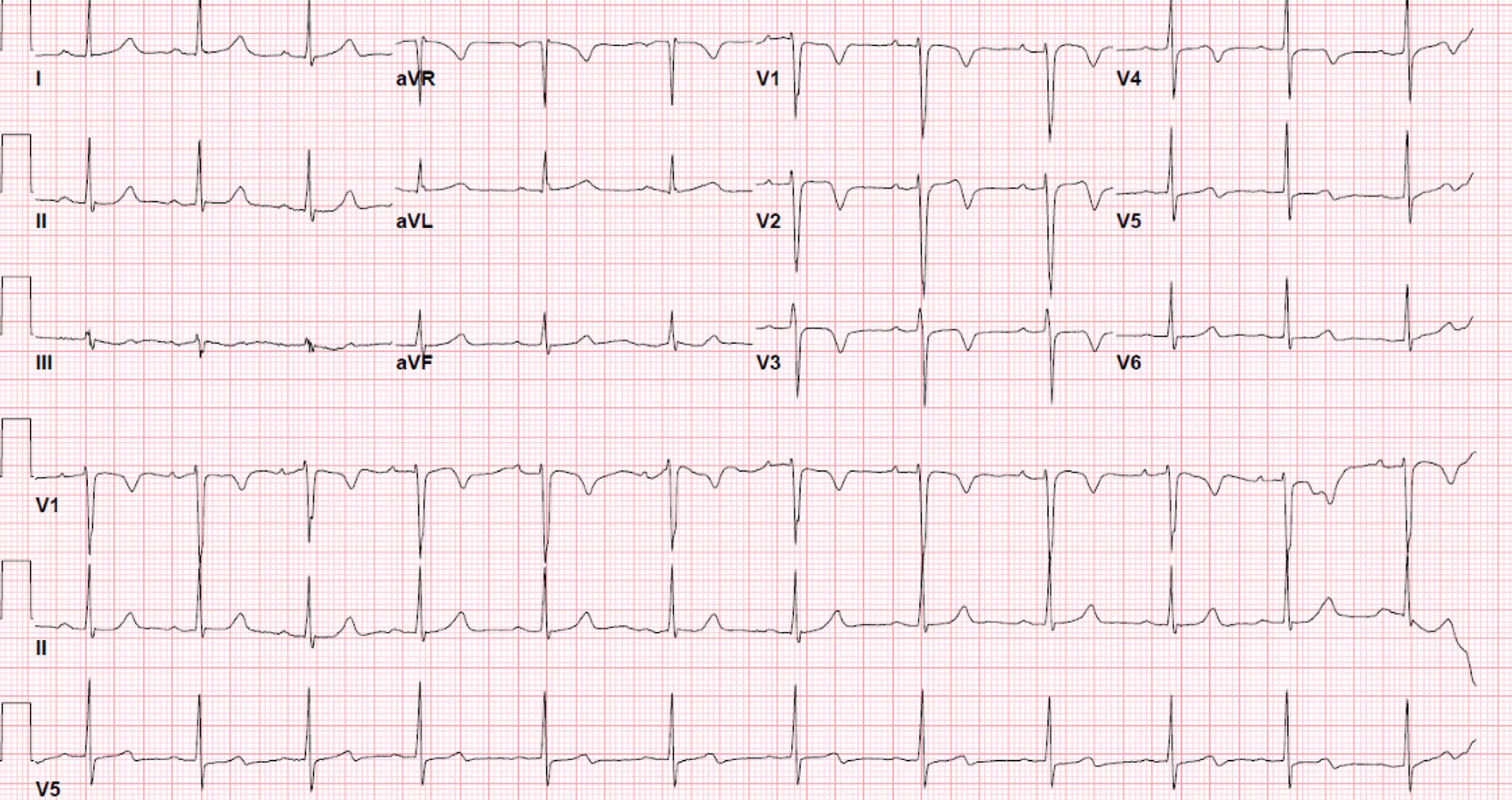
Electrocardiogram: T inversions in leads V1-V3 at restStress Echocardiogram: Hypokinesia at the anterior segment with dynamic ST-downsloping depressions at peak exercise, associated with breathlessness and vague chest discomfort2D Echocardiogram: Ejection fraction 56% with regional wall motion abnormalities at the anterior basal to mid segment, valves are normal.Troponin T levels 540pg/ml (normal < 14pg/ml)
ECHO2.avi

Relevant Catheterization Findings

Invasive coronary angiogramLeft Main Stem (LMS): Critical 99% distal LMS stenosis caused by calcified noduleLeft Anterior Descending (LAD): Severe 90% ostial calcified stenosis, severe 95% mid LAD stenosis, heavily calcified, with large coronary aneursyms at proximal and mid segmentsLeft Circumflex (LCx): Diffuse calcified disease non-dominant vesselRight Coronary Artery(RCA): Critical 99% stenosis caused by calcium nodule at mid RCA
Impression: Severe calcified 3 vessel disease
LMS pre.avi
LMS pre2.avi
RCA pre.avi

Interventional Management

Procedural Step

Heart team meeting was held, and patient outright refused bypass surgery due to age-related peri-operative risks and background COPD. Thus high-risk multivessel angioplasty was done in two stages.First procedure was angioplasty to RCA. Via femoral access ,and with temporary pacing wire for backup, engaged a JR 3.5 7Fr, workhorse wire then exchanged for rota-floppy wire and performed Rotational Artherectomy at 180000rpm.Successful procedure and stented with 3.5x38mm DES, NC balloon 4.0mm at high pressures, guided by intravascular ultrasound (IVUS)Second procedure was done after a week, again via femoral access, EBU 3.5 7Fr catheter engaged to LMS. Careful wiring with two workhorse wires into LAD and LCX, then predilated with 2.0x12mm NC balloon to open a small channel to provide flow. Optical coherence tomography (OCT) done showing severe stenosis of distal LM (MLA 1.4mm2) caused by eruptive calcium nodule and severe circumferential calcified plaque and mid LAD, with a OCT-Calcium score of 4/4, needing artherectomy. Decided to proceed withRotational Artherectomy with 1.5mm burr at 180000rpm. Guide manipulation and slower burr speed at 160000rpm used to improve wire bias and attain better debulking results. Successfully stented 3.0x28mm at mid LAD overlap with 3.5x23mm stent from proximal LAD to body of LM, NC 3.5mm mid LAD and POT at LMS with 4.5x8mmballoon at high pressure. Final OCT showed well expanded stent (expansion index145%) with good MSA and no stent malopposition.

RCA post.avi
LMS post.avi
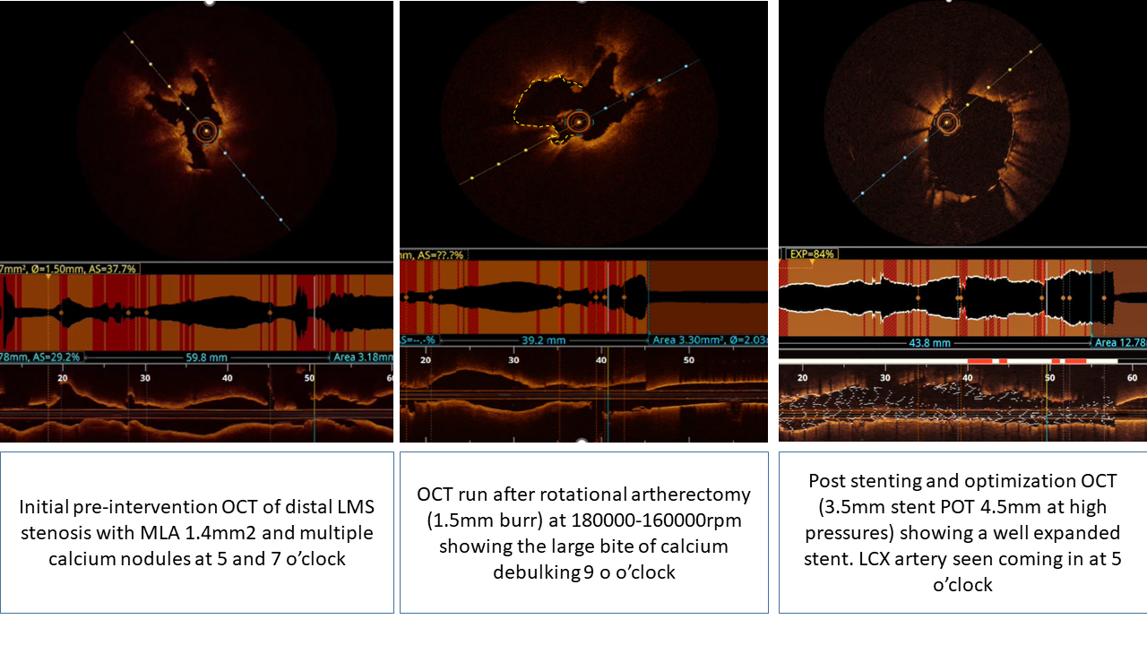
OCT 1st run before Rota.avi
OCT 2nd run after Rota.avi
OCT 3rd run after stent and optimization.avi

Case Summary

Patient recovered well post angioplasty and was discharged home the next day. He is well at 1 months clinic follow up with NYHA class 1 and no chest pain, and has returned to his usual routine of cycling. In conclusion, high-risk complex angioplasty can be successfully performed with low rates of complications when done with proper planning and in stages. This case illustrates how different intracoronary imaging modalities (IVUS and OCT) have been instrumental in choosing the right calcium debulking methods, ensuring good stent expansion and reducing the risk of stent failure. In summary, intracoronary imaging should be mandated in cases with heavily calcified plaque.